Story
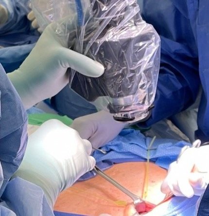
The hANDY-i is a portable imaging device for intraoperative vision assistance. Its dual RGB/NIR cameras capture information invisible to the naked eye, and assist surgeons to make better decisions, improving surgical outcomes.
Compared to existing technologies, the hANDY-i can display real-time structural and perfusion tissue information of parathyroids without interfering with the surgical workflow. All the operating room (OR) lights can remain on, and the surgeon can use the device whenever the need arises. This better visualization empowers surgeons to focus their attention on treating the patient efficiently and safely, without worrying about the costly and subjective risks that come from a dye injection or naked eye identification.

Our Technology, the hANDY-i
The hANDY-i is at the bedside, ready to be used whenever it is needed. Continuous, non-invasive visualization gives the surgeon the ability to treat the patient with confidence.
Faster and more accurate visualization of the parathyroids will decrease the total operating time. Patients can go home to their loved ones even faster now.
Dye free perfusion assessment will be seamlessly integrated into the clinical workflow, giving surgeons critical information in real-time.
We recently showed 94% device identification accuracy in early feasibility studies at Johns Hopkins Hospital¹
We are accelerating and innovating our technology both in benchtop and clinical testing. Our group was one of the first to show successful feasibility of machine learning and deep learning applications to parathyroid visualization¹

